Arabinogalactan and lipoarabinomannan
Mycobacteria are commonly classified as Gram-positive, although their cell wall remains impermeable to crystal violet without heating or phenol pre-treatment. This resistance to the penetration of dyes results from a dense barrier of fatty acids called mycolic acids, which are covalently bound to the polysaccharide arabinogalactan. The cell wall of mycobacteria contains a complex network of glycoconjugates, some of them being specific to the mycolata taxon. These include arabinogalactan, phosphatidylinositol-mannosides (PIM), lipomannan, and lipoarabinomannan (LAM). Besides Man, GlcNAc and MurNAc, the dominant monosaccharides found in the mycobacterial cell wall are the furanoside forms of Gal and Ara, abbreviated Galf and Araf.
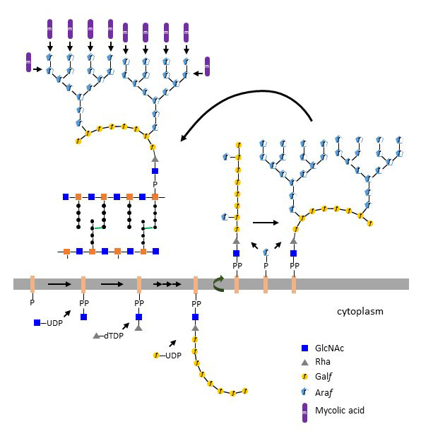
Figure X. Biosynthesis of arabinogalactan on decaprenol-PP and transfer to peptidoglycan in Mycobacterium spp.
Arabinogalactan is a large polysaccharide that is linked to peptidoglycan at its reducing end and carries multiple units of mycolic acid at its termini. The core of arabinogalactan is assembled at the cytoplasmic side of the cell membrane by first transferring GlcNAc to decaprenol-PP. This initial step is identical to the beginning of teichoic acid biosynthesis and dolichol-PP-oligosaccharide assembly in eukaryotes. The isoprenoid carrier in mycobacteria is however decaprenol (C50) and not undecaprenol (C55) like in other bacteria. After addition of α1-3 Rha to decaprenol-PP-GlcNAc from the donor dTDP-Rha, a chain of about 30 Galf is built up at the cytoplasmic leaflet by at least three Galf-transferases. The first enzyme transfers β1-4 Galf to Rha and the following ones transfer β1-5 Galf and β1-6 Galf alternately. The galactan polymer then translocates to the extracellular side of the membrane in a reaction probably mediated by an ABC-transporter. At the outer leaflet of the membrane, a series of Araf-transferases add two to three branched Araf trees each consisting of about 20 monosaccharides to each galactan chain. These Araf-transferases are embedded in the cell membrane and use decaprenol-P-Araf as donor substrate. Completed arabinogalactan units are then conjugated at their reducing end to MurNAc residues on peptidoglycan. Finally, several mycolic acid molecules are attached covalently to the Araf termini of arabinogalactan. The biosynthesis of mycobacterial peptido¬glycan proceeds as in other bacteria but decaprenol-PP is used as carrier instead of undecaprenol-PP.
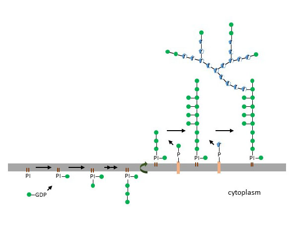
Figure X. Biosynthesis of lipoarabinomannan on phosphatidylinositol.
Whereas arabinogalactan covalently connects peptidoglycan and the mycolic acid layers, lipoarabinomannan is anchored in the cell membrane and in the mycolic acid layer through phosphatidylinositol. Phosphatidylinositol-mannosides, lipomannan, and lipoarabinomannan share the same initial biosynthetic steps, but only the biosynthesis of the more complex lipoarabinomannan will be addressed here. The first four to six Man units are added on phosphatidylinositol by cytoplasmic α1-2 and α1-6 Man-transferases using GDP-Man as substrate. The resulting glycoconjugate then flips to the outer leaflet of the cell membrane, where other Man-transferases using decaprenol-P-Man add about 20 monosaccharides. The resulting lipomannan is further extended by transfer of a branched Araf structure of about 50 to 80 monosaccharides. The lipoarabinomannan of pathogenic mycobacteria, such as Mycobacterium tuberculosis and Mycobacterium leprae, is capped by Man mono- or disaccharides, which are important for the binding and invasion of the host cells through Man-binding lectins. Arabinogalactan and lipoarabinomannan are essential for Mycobacterium viability. Truncated polysaccharides destabilize the barrier function of the cell wall and render bacteria more susceptible to antibiotics and environmental stress. In the context of pathogenic mycobacteria, lipoarabinomannan also contributes to immune evasion by suppressing the release of pro-inflammatory cytokines by dendritic cells and macrophages.
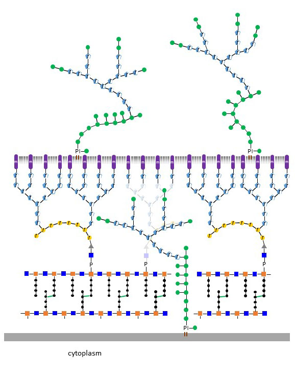
Figure X. Representation of the Mycobacterium cell wall with phospholipids and lipoarabinomannan embedded in the mycolic acid layer.
Various antibiotics target the mycobacterial cell wall, although the dense mycolic acid layer prevents the penetration of hydrophilic large molecules. Amphomycin blocks decaprenol-P-Man formation and prevents the elongation of phosphatidylinositol-mannosides after translocation to the extracellular side of the cell membrane. The action of amphomycin is not restricted to mycobacteria since this antibiotics also inhibits undecaprenol-PP-MurNAc-peptide translocation for peptidoglycan formation and also impairs dolichol-P-Man biosynthesis in eukaryotes. Other antibiotics, such as ethambutol, isoniazid and delamanid, are more specific to the mycobacterial cell wall. Ethambutol inhibits Araf-transferases, while isoniazid and delamanid inhibits mycolic acid biosynthesis.